Advancements in imaging technologies have significantly enhanced the precision and depth of preclinical oncology research. Optical luminescence and fluorescence imaging are two pivotal techniques employed to monitor disease progression and evaluate therapeutic efficacy. While they share several methodological similarities, their distinct mechanisms and applications render them uniquely valuable in different contexts.
Optical Luminescence Imaging
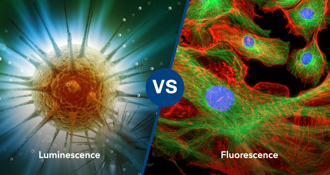
Optical luminescence imaging relies on the emission of light resulting from chemical reactions within biological systems. This technique is particularly advantageous for in vivo studies, offering high sensitivity and the capability to non-invasively track biological processes over time.
Key Characteristics:
- High Sensitivity: Capable of detecting minimal light emissions, facilitating the observation of subtle biological changes.
- Non-Invasive Monitoring: Allows for the continuous, non-invasive observation of live animal models.
- Versatility: Applicable to various research domains, including tumor growth tracking, gene expression analysis, and infectious disease studies.
Fluorescence Imaging
Fluorescence imaging utilizes fluorescent molecules that emit light upon excitation by specific wavelengths. This method is widely adopted for its high specificity and ability to simultaneously visualize multiple targets within a sample.
Key Characteristics:
- High Specificity: Enables the detailed visualization of specific proteins, cells, or subcellular structures.
- Multiplexing Capability: Allows for the concurrent imaging of multiple targets using different fluorophores.
- Quantitative Analysis: Provides quantitative measurements of the distribution and abundance of fluorescently labeled entities.
Comparative Overview
Despite their differences, optical luminescence and fluorescence imaging share fundamental similarities:
- Non-Invasive: Both techniques enable the non-invasive examination of biological processes in vivo.
- High Sensitivity: Both methods are adept at detecting low levels of light emission, making them suitable for studying minute biological changes.
- Broad Applicability: Both imaging modalities are versatile and widely used across various research areas, including oncology, infectious diseases, and gene expression studies.
Distinctive Features:
- Mechanism of Light Emission: Optical luminescence is driven by chemical reactions, whereas fluorescence relies on the excitation of fluorophores.
- Signal Duration: Luminescence signals tend to have a longer duration, supporting extended imaging sessions. Fluorescence signals, however, generally decay more rapidly.
- Background Noise: Luminescence imaging often exhibits lower background noise compared to fluorescence, which can be influenced by tissue autofluorescence.
Application in Preclinical Oncology: TD2’s Imaging Capabilities
At TD2, we harness the power of both optical luminescence and fluorescence imaging to deliver comprehensive preclinical evaluation services. Our imaging technologies, exemplified by the Spectral Instruments Imaging AMI HTX, enable precise monitoring of tumor growth kinetics and therapeutic responses in orthotopic tumor models.
Key Features of TD2’s Imaging Services:
- High-Quality Imaging: Our equipment supports both optical luminescence and fluorescence imaging, offering high-resolution images essential for accurate tumor assessment.
- Advanced Data Analysis: Equipped with sophisticated software, our systems provide detailed illustrations of drug progression in longitudinal studies.
- Smart Exposure Technology: Ensures the capture of both faint and strong signals within a single image, optimizing sensitivity and clarity.
For a deeper understanding of our imaging capabilities and their potential applications in your research, please visit our Imaging Services page.